Project Methodology

- 1. Methodology Introduction
- 2. Cell Biologist
- 3. 1758 Military Dock
- 4. Scanning Electron Microscope
- 5. Amoeba Cement
- 6. Centropyxis
- 7. Side View of a Centropyxis
- 8. A Difflugia Amoeba
- 9. Side View of Difflugia
- 10. View of Difflugia Aperture
- 11. Difflugia and Substrate
- 12. Difflugia Tail
- 13. Tiled Drawings and Micrographs
- 14. Nanolithography
- 15. Silicon Wafer
- 16. Amoeba Gold
- 17. Underwater Easel
- 18. Art Exhibit on the Lakebed
- 19. Underwater Diorama
- 20. Art Gallery
- Related exhibit:The Search for Underwater Sites from the French and Indian War
- © 2009 T Kurt Knoerl Last updated 19 July 2009
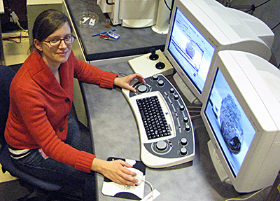
Scientific Artist Elinor Mossop Views the Magnified Images of the Testate Amoebae (Photo Courtesy Sam Bowser)
A Scanning Electron Microscope (SEM) directs an extremely thin beam of electrons across a specimen in a raster pattern. At each point along the scan, the energy produced by electron bombardment on the surface of the specimen is detected and displayed on a screen, building up an image point-by-point. Information about the topology of the specimen, as well as its conductivity and elemental composition, is thus revealed at nanometer resolution.
View more information on SEMs here.
Previous Next